
: Admin : 2022-03-05
How is the gastrointestinal tract formed?
Human beings have well developed, complex, highly evolved organ systems, assembled internally, in a distinct, specific position and method within the thoracic- abdominopelvic cavity. The heart and lungs being the supreme organs of paramount importance are guarded within the thoracic cavity. The abdominal cavity is located between the thoracic and pelvic cavities. The abdominal cavity is the largest hollow sac, filled with the stomach, liver, pancreas, intestines and gall bladder. Rectum, uterus, urinary bladder lie in the lower part of the abdominal cavity. Kidneys lie in the posterior pelvic region. The entire abdominal cavity is enclosed in the peritoneum. It is a membranous structure that covers the inside wall of the abdominal cavity. This is called the parietal peritoneum. It also acts as a shield, covering all organs lying within the abdominal cavity. This is called the visceral peritoneum that protects the abdominal viscera. All organs are attached firmly by means of the peritoneum which also divides the abdominal cavity into distinguished compartments. Anatomically, it is divided into eight quadrants. Every quadrant encloses organs that together form the digestive system. The mesentery is folds of the peritoneum that are attached to the walls of the abdomen. Omentum is a term given to the peritoneum that surrounds nerves, blood vessels and lymph vessels that lie in the abdomen.
The abdomen is tightly packed with organs that aid in the function of the digestive system. Development of the digestive tract takes place at about five weeks of pregnancy in an embryo, inside the womb of the mother. At about five to seven weeks, a layer of cells rolls into a tubular structure that expands to form the foregut, midgut and hindgut. The foregut shapes into the oesophagus, stomach, liver and pancreas. At about ten to twelve weeks of pregnancy, midgut develops into intestines. Within nine to eleven weeks, the hindgut develops to form the anus and rectum. Soon, by twenty-three weeks of pregnancy, bowel movements are observed within the child's gut or digestive system. As the foetus is born, grows into a child and later into an adult, the digestive system that begins from the mouth and extends to the anus, aids in the process of mastication, digestion, absorption and assimilation of a bolus of food. Every organ has a vital role to play in digestion. Food reaches the stomach through a narrow tube called the oesophagus. Digestive juices or bile from the liver and gall bladder break down fats, removing toxins. The pancreas produces digestive enzymes that break down proteins, fats and carbohydrates. The partially broken down bolus of food then enters the intestines. Here it is further broken down, absorbed as nutrients and made to solidify into waste products. The twenty feet long small intestine absorbs essential nutrients and water. The large intestine is about five feet long. Its main function is to absorb water from waste forming stool. The stool now passes into the rectum. The absorption of water, nutrients that create dry waste stool, its passage into the rectum stimulate nerves that create an urge to defecate.
Hence every part of, the organ forming the digestive tract is very important for the metabolism and growth of human beings. A disease or any pathology within the digestive tract can disturb the functions of several other organ systems. Diseases of the digestive tract affect the external peritoneum, organs of the abdominal cavity and surrounding structures. Although the peritoneum forms a layer around the abdomen and its organs, it is the foremost tissue to be affected or damaged by any pathological condition or state present in the abdomen. A hernia is a disease that not only harms the vital organs of the abdomen but also the peritoneum causing distress to a patient’s health.
What is a Hernia?
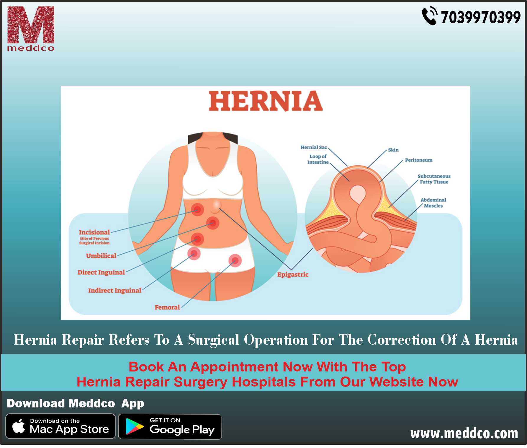
The muscles and tissues surrounding the abdomen weaken as age advances in some people. Some people are born with weak muscles or have inherited traits that cause musculature, fatty tissue to loosen, protrude out of the abdominal wall or cavity, obstructing functions of surrounding healthy tissues and organs. A hernia is the abnormal protrusion or exit of a tissue or an organ such as an intestine through or outside the abdominal wall. The connective tissue, muscle or fascia become weak from pressure, in patients who are obese, or patients born with weak muscles, this causes an organ to squeeze out through this weak spot that is medically termed as a hernia. A gap or opening is formed from excessive pressure in the muscular wall of the abdomen that forms a bulge as contents protrude outside its cavity. In most of these hernias, peritoneum also bulges outside the abdomen along with parts of an organ. Hernias are commonly found around the belly, umbilicus and near groin areas.
What are the different types of Hernias?
Hernias are caused mainly by pressure and weaknesses of muscles surrounding abdominal organs. Chronic diarrhoea, constipation, lifting heavy objects or weight, persistent sneezing or coughing, obesity, poor nutrition and smoking weaken abdominal muscles, which causes herniation or protrusion of organs packed inside the cavity. The classification of hernia is based on the part or area from where a bulge or opening through which an organ exit are found on the abdominal wall.
INGUINAL HERNIA – Incidence of inguinal hernia is observed in males due to natural weakness in the groin area. About ninety-six per cent of all groin hernias are inguinal where intestines or bladder protrude through the abdominal wall into the inguinal canal or groin region. It is a very common clinical pathology that needs surgical treatment procedures.
INCISIONAL HERNIA – An intestine pushes its way through the abdominal wall from the area where a previous surgery was done having scar formation near the site of the incision. It is common in elderly patients or obese patients whose abdominal muscles become weak or dysfunctional, inactive from previous surgery.
FEMORAL HERNIA – This hernia is most common in females who are pregnant and overweight. Intestines enter the canal carrying the femoral artery, into the upper thigh.
UMBILICAL HERNIA – A part of the small intestine herniates through the abdominal wall near the umbilicus. It affects women who are obese or have given birth to many children. It is also seen commonly in newborn infants who have inherited weak abdominal muscle walls.
HIATUS HERNIA – A hiatus is an opening in the diaphragm through which the oesophagus passes. The upper part of the stomach squeezes through this opening causing a hiatus hernia.
They are also classified according to the extent or spread of protruded organs outside the abdominal wall.
REDUCIBLE HERNIA – These types can be pushed back into the opening through which it formed a bulge and developed a hernia.
IRREDUCIBLE HERNIA – When organs or the tissues that protrude through the opening, cannot be pushed back through, it is called an irreducible hernia.
STRANGULATED HERNIA – The blood supply to the hernia gets obstructed or cuts off when an organ or tissue gets struck or strangulated inside the hernia.
DIRECT HERNIA occurs develops in adulthood, from pressure or weight, when walls of abdominal muscles become weak. Intestines push their way through this weak musculature, forming hernia. INDIRECT HERNIA is seen during infancy. The inguinal ring is an area of abdominal muscle tissue that fail to close when the baby is inside the mother's womb. When the ring remains open, parts of the intestines bulge out causing a hernia.
What are symptoms of Hernia?
When a patient suffers from a hernia, he or she feels different symptoms. Asymptomatic hernias are also accidentally found during diagnostic procedures. They are silent, present within the abdominal sac, which in later years may cause symptoms that need medical intervention.
Swelling beneath the skin of the abdomen or groin can be felt by the patient and treating physician. The patient feels heaviness in the abdomen with constipation and bloody stools. Pain and discomfort can be felt by patients while bending or lifting weight. Weakness, pressure in the groin is often felt in the case of inguinal, femoral, incisional and umbilical hernias. Vomiting with heartburn is felt by patients who suffer from hiatus hernia. Strangulation of hernia is the most common complication if surgical treatment is not done. If a patient feels these symptoms, medical advice or consultation is crucial to save the life of the patient and prevent complications like infections, nerve damage, fistula, urinary tract infections and haemorrhage.
A doctor examines the patient, records the signs and symptoms of patients. An ultrasound and X rays are common diagnostic tests to locate the site of the hernia. Abdominal MRI scan, CT scan and endoscopy, are also diagnostic procedures that may be advised by the surgeon. Surgery is often a choice of treatment for hernias. However, a medical professional or a surgeon decides on the treatment protocol, once he concludes the final assessment and diagnosis.
What is HERNIORAPPHY?
HERNIORAPPHY is the surgery done to repair a hernia. The surgeon repairs the weakness around the abdominal wall. Surgical repair of a hernia is termed HERNIORAPPHY.
Direct hernias that show bulging of abdominal wall, the surgeon pushes back the contents back to their normal anatomical position. The weak muscle spot is then sutured together through the edges of healthy muscle tissues. After the muscle gets repaired, a mesh is implanted over it to reinforce and strengthen the muscle area to prevent the future incidence of hernia. This is termed hernioplasty.
Surgery is done following certain definite steps.
General or local anaesthesia is given to the patient depending upon the extent of herniation or it is associated with any complications. The area is cleansed, sanitized before an incision is made on the abdominal wall. An incision is then made parallel to the area adjacent to the inguinal ligament. The hernia sac with all the contents that are bulged, is felt, identified and pushed back to its original position. Peritoneum is that bulges out from the sac is also placed or resected back to its anatomical position.
When a small sac of hernia protrudes out containing lesser contents, then the abdominal wall is stitched back, through HERNIORAPPHY surgery, after each structure and tissue is placed back to its normal position to restore and revive its physiological function. If a larger area is affected, a mesh is implanted by the surgical procedure called hernioplasty. The initial cut or incision on the abdomen is sutured or closed back and dressing with an antiseptic solution is applied over the area. Recovery after surgery takes about three weeks after HERNIORAPPHY surgery is performed. Patients are advised not to lift heavyweight. Pain and swelling may be felt near the area of the incision. Ice or cold pack can be applied for fifteen minutes to heal pain and swelling.
HERNIORAPPHY a the best surgical treatment procedure that treats patients with hernias. Laparoscopic surgery is also an advanced treatment option using cameras and equipment, which is a minimally invasive procedure. Robotic-assisted hernia repair is also yet another treatment procedure. However, surgeons still opt for the traditional method-- HERNIORAPPHY as a better surgical treatment procedure.
How can the future incidence of hernia be prevented?
Hernias can recur in one out of ten patients from every hundred open HERNIORAPPHY surgeries done. Postoperative care is of prime importance. Children need more care and attention post-surgery. For pregnant females who have a hernia before or during pregnancy, a HERNIORAPPHY surgery is advised with utmost care post the procedure is performed to prevent infections and complications at the time of delivery. A Cesarean section during delivery leaves the muscles around the scar or an incision weak. Care should be taken by the surgeon that such incisions are sutured back in such a way that incisional hernia can be prevented. Children born with weak muscles should be under observation by the surgeon to avoid an umbilical hernia. Prompt medical care should be taken to diagnose this condition, the area around the weak muscles should be repaired, tightened to avoid the risk of a child developing a hernia.
Hernias can be prevented by maintaining ideal body weight. A healthy diet, lifestyle and regular exercises help to maintain body weight in order to avoid obesity. Fruits, vegetables and adequate water intake prevent constipation. Pressure on the abdomen from lifting heavy weights should be avoided. Smoking should be stopped, which induce coughing in patients that may trigger hernias to prevent its recurrence. Yoga helps patients to cope, adopt to a healthy, balanced lifestyle. A physical therapist may guide the patients on exercises that strengthen abdominal fats and muscles.
hernia hernioropphy abdomin repair surgery treatment
No Comments